Research Article - (2021) Volume 12, Issue 12
Fracture Resistance of Anterior Endocrown vs. Post Crown Restoration an In-vitro Study
Manar al-Fadhli1*, Cherif Mohsen1 and Hesham Katamich2Abstract
Objective: The purpose of this study is to study the fracture resistance of anterior endocrown versus post crown restoration.
Materials and methods: Thirty caries free of extracted human maxillary canine with complete root formation were selected for this study. Also two types of ceramics were used: IPS Emax press and Celtra Press. Thirty samples were fabricated and divided into 2 groups (15 samples each) according to the type of ceramic used. Then each subgroup was subdivided into 3 subgroups (5 samples each) according to the type of restorations: Endocrown 6 mm, Endocrown 10 mm, post+core+crown. All specimens were subjected to a thermocycling procedure in automated thermocycling machine. Samples were thermocycled for 5000 cycles, between 5°C-55°C with a dwell time 15 seconds. All samples were individually mounted on a computer controlled materials testing machine for a fracture resistance test. After fracture resistance test, all specimens in the test groups were viewed using a USB digital-microscope, magnification × 35, and the images were captured and transferred to a IBM personal computer equipped with the Image-tool software (Image J 1.43 U, National Institute of Health, USA) to determine failure mode pattern according to the following categorization.
Results: Regarding the fracture resistance testing, the results of the present study revealed that there was no statistically significant difference between mean fracture resistance values of IPS Emax press (235.54 N) and Celtra press; (244.98 N) where both showed statistically non-significant highest mean fracture resistance.
Conclusion: The results obtained in this study revealed a non-significant difference between the endocrown restorations and the post and core and crown. Although endocrown showed higher fracture resistance values compared to the post and core and crown. Also the results showed non-significant difference between the endocrown restorations with different designs. Although endocrown (10 mm) showed higher fracture resistance values than endocrown (6 mm) which may be due to an increase in surface area.
Keywords
Canine teeth, Endocrown, Endodontically treated teeth, Fiber post, Emax, Celtra
Introduction
Endodontically treated teeth restoration is compromised primarily because increased risk of coronal destruction that results in tooth fracture during function. Before the introduction of adhesion in dentistry, the coronal restoration endodontially treated teeth has been mainly performed with metallic and macro mechanically retained posts. In the past, a length of post equal to three fourths of the root canal or at least equal to the crown length (Goodacre CJ, Spolnik KJ, 1995; Abramovitz L, et al., 2001). A restorative option for endodontically treatment an endocrown is. It consists of a full crown that extends a post into the pulp chamber and/ or pulp canals as one unit (Lander E and Dietschi D, 2008). One of option to restore severe destructed, supragingival structure of posterior teeth (Stockton L, et al., 1998) endocrown. These restorations are the most prevalent in case of damaged molars crowns, narrow and short roots, obturated canals or limited interocclusal space (Güngör MB, et al., 2017). The endocrown are mechanically anchored in pulp chambers (3-4 mm element) (Güngör MB, et al., 2017; Reich S, et al., 2004; Mannocci F and Cowie J, 2014) and strongly adhesively bonded with hard dental tissues using resin cements (Reich S, et al., 2004). The little dental structures preparation when compared with conventional post and cores (Mannocci F and Cowie J, 2014), as well as lack of intervention in the root canals (Edelhoff D, Sorensen J, 2002). They need less time to be made and less interfaces between part of the restorations and the teeth Compared to traditional methods.
One of ZLS classes, celtra press (Dentsply Sirona) is a new material that can deliver the highest level of esthetics mimicking natural teeth based on having an amazing chameleon effect, perfect balance of translucency and natural like opalescence only by polishing the material after pressing it (Celtra press Fact File, 2017). Feld spathic ceramic is the most material for Endocrown (e.g. Vitablocs Mark II, Vita Zahnfabirk, Bad Säckingen, Germany and Cerec Blocks, Sirona Dental Systems, Bensheim, Germany), leucite ceramic (e.g. IPS Empress CAD, Ivoclar Vivadent Schaan/ Liechtenstein), lithium disilicate ceramic (e.g. IPS Emax Press, Ivoclar/Vivadent, Schaan/Liechtenstein) or resin composite (e.g. Lava Ultimate, 3 M ESPE, St Paul, MN, USA) (Dietschi D, et al., 2007). Ceramic endocrown are made by sintering, pressing or more often CAD/CAM technology. The restorations are aesthetically attractive.
The fracture resistance of ETT has been reported to be mainly dependent on the amount of adhesive surface, the amount of the remaining tooth structure and the quality of adhesion (Garbin CA, et al., 2010). The way of a post in the retention of the core material is relevant for posterior teeth where masticatory loads are essentially compressive (Reeh ES, et al., 1989). in vitro studies on adhesive restoration tested to analyse fracture strength and mode of failure of cuspid endodontically treated teeth (Zarow M, et al., 2009).
Materials and Methods
The Thirty caries free of extracted human maxillary canine with complete root formation no crack or fracture. Teeth were cleansed of calculus and soft tissues, with dental scalers, nylon bristle brushes, and pumice with low speed hand piece. They were stored in 5% sodium hypochlorite for 15 minutes at room temperature for sterilization of extracted teeth.
All teeth were mounted in epoxy resin block using a custom-made round Teflon shape sample holder (2 cm length, 2 cm internal diameters) in a vertical direction using centralizing device. The amount of mixed material that the application requires was determined according to manufacture structure.
The crowns of teeth were cut perpendicular to the long axis 2 mm above the cemento-enamel junction from the proximal surface using a super coarse diamond disc and copious irrigation to avoid dryness (Figure 1).
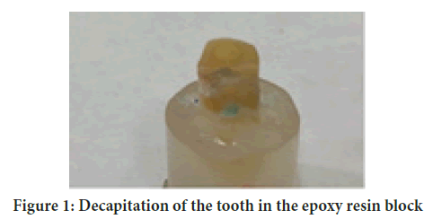
Figure 1: Decapitation of the tooth in the epoxy resin block
The access cavity preparation was done including completely pulp chamber de-roofing and orifice was exposed. Then the radicular pulp was extirpated with a barbed broach.
The working length of the canal was determined by inserting a size 10 K-type file into the root canal terminus. Then a glide path was performed with a size 15 K-type file. The working length was demonstrated by the initial peri-apical x-ray and kept 0.5 to 1 mm away from the apical constriction. The filling process is continued by using 20 then 25 sized files respectively. The root canal was instrumented with Pro-Taper Universal instruments in a sequence of (SX, S1, S2, F1, F2) with an adjustable-torque motor* (NSK, Japan). The root canal was irrigated with 2 ml 1% sodium hypochlorite solution after each instrument change. After the instrumentation, the canal was flushed with 5 ml 17% EDTA and 5 ml sodium hypochlorite and then dried with paper points.
The master gutta percha cone inserted into the canal for proper working length insurance. The obturation process was done in the help of Adseal sealer. Adseal is resin based root canal sealer which is a paste type of dual syringe. It provided easy and standard mix of quit volume units (2:1 wt. Ratio) of base and catalyst according to manufacture instructions. They were mixed together on a mixing pad using spatula for 15-20 seconds until creamy homogenous mix was gained. The sealer was applied to the cone size F2 and inserted into the canal. A hot burnisher was used to remove the protruded gutta percha.
Classification of samples
Thirteen were divided into two main groups according to the type of restoration and three subgroups according to the designs of preparation (Table 1):
| Group (A) LDS (15 samples) |
Subgroup (A1) | Post+core+crown | Samples |
| Subgroup (A2) | Endocrown 6 mm |
5 samples | |
| Subgroup (A3) | Endocrown 10 mm |
5 samples | |
| Group (B) ZN+LDS (15 samples) |
Subgroup (B1) | Post+core+crown | 5 samples |
| Subgroup (B2) | Endocrown 6 mm | 5 samples | |
| Subgroup (B3) | Endocrown 10 mm | 5 samples |
Table 1: Classification of the samples
Conventional post and core supported crowns group preparation
For each tooth, post space preparation began with the removal of gutta percha to a depth of 10 mm from the coronal tooth structure using gates glidden drills, then Glassix radiopaque glass fiber composite post drill* was used to remove any residual root filling and complete canal preparation with water spray coolant and at a low speed (30000 R.P.M). Each glass fiber post was reduced to a length of 12.0 mm by cutting the coronal end with a diamond separating disc under copious coolant, resulting in a dowel extending 2 mm above the coronal surface of the trimmed crown.
The root canal was etched with 35% phosphoric acid** (Where ** is Scotch bond etchant, 3m ESPE Dental products) for 15 seconds, washed with water spray, and then the excess water was removed from the post space using paper points.
RelyX unicem dual-cure self-adhesive resin cement* (Where * is Nordon, swiss dental products of distinction) was used for luting of glass-fiber poste.
After post cementation the remaining tooth structure was etched followed by the application of adhesive bonding agent according to manufactures instruction. Composite was applied with increments to the tooth structure and polyester crown forms for standardizing the core height, then light activated for 40 seconds for each surface. Axial walls were prepared with a circumferential 90° shoulder margin 1 mm wide with rounded internal line angles, located on sound tooth structure 1 mm from the cemento-enamel junction. And with a 10° convergence angle using diamond stone.
A special milling machine was used for teeth preparation. The assembly incorporate a conventional-speed 30.000 rpm straight hand-piece perpendicular to the surveyor platform.
Preparation design of endocrown: The entrance to the pulpal canal was opened and gutta percha is removed to a depth 6 mm and without undercuts in the access cavity. A cylindrical-conical diamond bur was used to make the coronal pulp chamber and endodontic access cavity continuous. It has a total occlusal convergence of 7°. The width and thickness of the enamel strip should be maintained by preserving as much tissue as possible from the root canal walls. The depth of the cavity should be 6 mm.
The pulp chamber was cleaned with ultrasound and its floor thoroughly. Orifice and undercuts of mesial and distal were protected using an adhesive system and flowable resin.
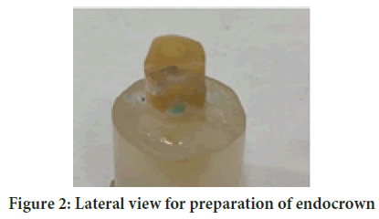
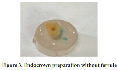
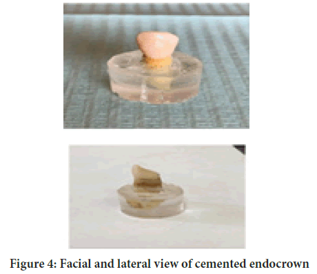
The butt joint, or cervical sidewalk, was the base of the restoration, and it provided a stable surface that could withstand the compressive stresses. The prepared surface was made parallel to the occlusal plane so that the stress resistance was ensured along the major axis of the tooth. The pulp chamber cavity ensured retention and stability. Extracoronally, the remaining vertical portion of the crown was prepared with a bevel labial and palatal and rounded internal line angles, located on sound tooth structure (Akkayan B and Gulmez T, 2002) (Figures 2-4).
Figure 2: Lateral view for preparation of endocrown
Figure 3: Endocrown preparation without ferrule
Figure 4: Facial and lateral view of cemented endocrown
Specimen's classification
The specimens will be divided into 6 sub-groups-
a) The first subgroup consisted of 5 specimens involved endodontically treated maxillary canine restored with glass fiber posts cemented with rely x cement, composite filling cores and conventional IPS Emax press crowns following the manufacture instructions.
b) The second subgroup consisted of 5 specimens involved endodontically treated maxillary canine and restored with IPS Emax endocrown according to design with 6 mm preparation.
c) Third subgroup consisted of 5 specimen involved endodontically treated maxillary canine and restored with IPS Emax Endocrown according to design with 10 mm preparation.
d) The forth subgroup consisted of 5 specimens involved endodontically treated maxillary canine and restored with glass fiber posts cemented with rely x cement, composite filling cores and celtra press ceramic crowns following the manufacture instructions (Mannocci F, et al., 1999).
e) The five subgroup consisted of 5 specimens involved endodontically treated maxillary canine and restored with celtra press Endocrown according to design with 6 mm preparation
f) The six subgroup consisted of 5 specimen involved endodontically treated maxillary canine and restored with celtra press Endocrown according to design with 10 mm preparation.
Thermocycling
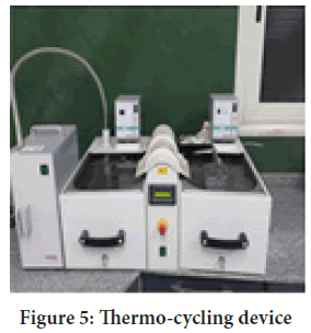
All specimens were subjected to a thermocycling procedure in automated thermocycling machine. Samples of retention test were thermocycled for 5000 cycles, between 5°C-55°C with a dwell time 15 seconds, but specimens of microleakage test the number of cycles used was 500 cycles between 5°C-55°C with a dwell time 25 seconds (Figure 5).
Figure 5: Thermo-cycling device
Test procedure (fracture resistance test)
Tests were performed using Bluehill Lite Software from Instron®.
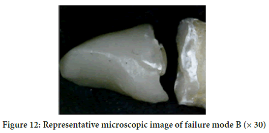
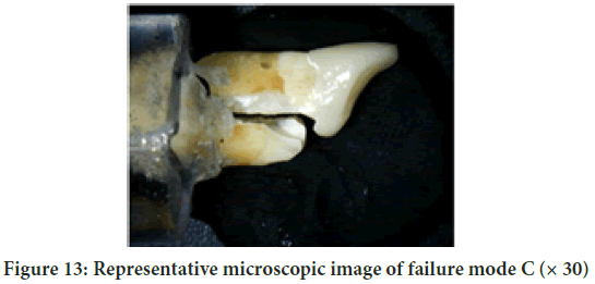
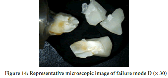
After fracture resistance test, all specimens in the test groups were viewed using a USB digital-microscope (U500 × Digital Microscope, Guangdong, China), magnification x35, and the images were capture and transferred to a IBM personal computer equipped with the Image-tool software (Image J 1.43 U, National Institute of Health, USA) to determine failure mode pattern according to the following categorization (Table 2).
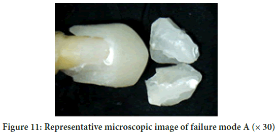
| Failure mode A | The lowest point of fracture line is located above cervical line |
| Failure mode B | The lowest point of fracture line is located between cervical line and tooth root embedded in resin |
| Failure mode C | The lowest point of fracture line is located inside tooth root embedded in resin |
| Failure mode D | More than one fracture lines run vertically and horizontally |
Table 2: Classification of failure modes
Statistical analyses
Data analysis was performed in several steps. Initially, descriptive statistics for each group results. One-way ANOVA followed by pair-wise Tukey’s post-hoc tests were performed to detect significance between groups. Student t-test was done between subgroups. Fisher exact test for qualitative data was done for mode of failure. Statistical analysis was performed using Grapg-Pad Instat statistics software for Windows (www.graphpad.com). p values ≤ 0.05 are considered to be statistically significant in all tests.
Data were presented as mean, standard deviation (SD), confidence intervals (low-high) for values. The results were analyzed using Graph Pad Instat (Graph Pad, Inc.) software for windows. A value of p<0.05 was considered statistically significant. After homogeneity of variance and normal distribution of errors had been confirmed, one-way ANOVA and student t-test was done for comparison between subgroups and main groups respectively. Two-way was done to detect effect of each variable (material and restorations). Chi square test was performed between failure patterns. Sample size (n=5/subgroup) was large enough to detect large effect sizes for main effects and pair-wise comparisons, with the satisfactory level of power set at 80% and a 95% confidence level.
Results
Fracture resistance
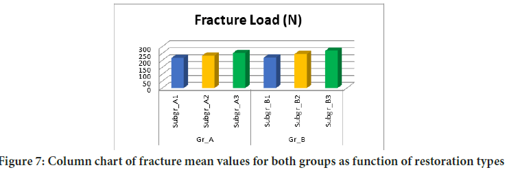
The mean values and standard deviation of fracture resistance (N) for both groups as function of restoration types are summarized in Table 3 and graphically drawn in Figures 6 and 7.
| Variable | subgroups | Mean | SD |
|---|---|---|---|
| Gr_A | Subgr_A1 | 218.01B | 21.13 |
| Subgr_A2 | 234.74B | 42.56 | |
| Subgr_A3 | 253.86B | 29.59 | |
| Gr_B | Subgr_B1 | 220.02B | 18.59 |
| Subgr_B2 | 245.54B | 35.39 | |
| Subgr_B3 | 269.39B | 45.34 |
Note: Different letter in the same column indicating statistically significant difference (p<0.05*: Significant (p<0.05) ns: Non-significant (p>0.05)
Table 3: Mean values ± SDs of fracture resistance (N) results for both groups as function of restoration types
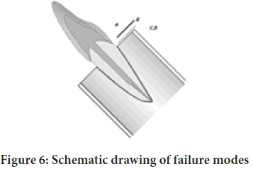
Figure 6: Schematic drawing of failure
Figure 7: Column chart of fracture mean values for both groups as function of restoration types
With Gr_A: There was not statistically significant difference (p<0.05) as Subgr_A3 recorded mean value (253.86 ± 29.59 N) followed by Subgr_ A2 (234.74 ± 42.56 N) while Subgr_A1 recorded the lowest statistically significant (p<0.05) mean value (218.01 ± 21.13 N) as indicated by one-way ANOVA test (p=0.002<0.05). Pair-wise Tukey’s post-hoc test showed non-significant (p>0.05) difference between Subgr_A1, Subgr_A2 and Subgr_A3 (Table 3 and Figure 6).
Group A vs. Group B
Subgroup 1: It was found that Subgr_B1 recorded statistically non-significant higher fracture resistance mean value (220.02 ± 18.59 N) than Subgr_A1 (218.01 ± 21.13 N) as indicated by student t-test (p=0.8771 >0.05) (Table 4 and Figure 7).
| Variable | Groups | Mean | SD | 95% CI | t-test | |
|---|---|---|---|---|---|---|
| Low | High | p value | ||||
| Subgr_1 | Gr_A | 218.01 | 21.13 | 191.77 | 244.24 | 0.8771 ns |
| Gr_B | 220.02 | 18.59 | 196.94 | 243.09 | ||
| Subgr_2 | Gr_A | 234.74 | 42.56 | 181.9 | 287.57 | 0.6742 ns |
| Gr_B | 245.54 | 35.39 | 201.6 | 289.47 | ||
| Subgr_3 | Gr_A | 253.86 | 29.59 | 267.12 | 340.6 | 0.1924 ns |
| Gr_B | 269.39 | 45.34 | 213.09 | 325.68 | ||
Note: *: Significant (p<0.05); ns: Non-significant (p>0.05)
Table 4: Comparison of fracture resistance (N) results (Mean values ± SDs) between both groups as function of restoration types
Subgroup 2: It was found that Subgr_B2 recorded statistically non-significant higher fracture resistance mean value (245.54 ± 35.39 N) than Subgr_A2 (234.74 ± 42.56 N) as indicated by student t-test (p=0.6742>0.05) (Table 4 and Figure 7).
Subgroup 3: It was found that Subgr_A3 recorded statistically non-significant higher fracture resistance mean value (253.86 ± 29.59 N) than Subgr_B3 (269.39 ± 45.34 N) as indicated by student t-test (p=0.1924>0.05) (Table 4and Figure 7).
Total effect of material on fracture resistance
Regardless to restoration type, totally it was found that Gr_A recorded statistically non-significant higher bi-axial flexure strength mean value (235.54 ± 31.09 N) than Gr_B (244.98 ± 33.11 N) as indicated by two way ANOVA test (p>0.05) (Table 5 and Figure 8). The failure modes of the different groups are listed in (Table 6) and (Figures 9-14).
| Variables | Groups | Mean | SD | Tukey’s rank | Statistics (p value) |
|---|---|---|---|---|---|
| Material group | Gr_A | 235.54 | 31.09 | A | 0.543 ns |
| Gr_B | 244.98 | 33.11 | A |
Note: Different letter in the same column indicating statistically significant difference (p<0.05*: Significant (p<0.05); ns: Non-significant (p>0.05)
Table 5: Comparison between total fracture resistance results (Mean values ± SDs) as function of material group
| Variable | Subgroups | Failure mode | Chi square test | |||
|---|---|---|---|---|---|---|
| A | B | C | D | P value | ||
| Gr_A | Subgr_A1 | 60% | 20% | 20% | 0% | <0.0001* |
| Subgr_A2 | 60% | 20% | 20% | 0% | ||
| Subgr_A3 | 0% | 20% | 20% | 0% | ||
| Gr_B | Subgr_B1 | 60% | 20% | 20% | 0% | <0.0001* |
| Subgr_B2 | 60% | 20% | 20% | 0% | ||
| Subgr_B3 | 20% | 20% | 20% | 0% | ||
| Chi test | p value | <0.0001* | ||||
Note: *: significant (p<0.05); ns: non-significant (p>0.05)
Table 6: Chi square test comparing between failure modes results for both groups as function of restoration types
Figure 8: Column chart comparing fracture mean values between both groups as function of restoration types
Figure 9: A column chart of total fracture resistance mean values as function of material group
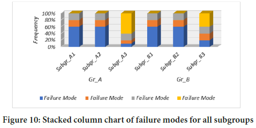
Figure 10: Stacked column chart of failure modes for all subgroups
Figure 11: Representative microscopic image of failure mode A (× 30)
Figure 12: Representative microscopic image of failure mode B (× 30)
Figure 13: Representative microscopic image of failure mode C (× 30)
Figure 14: Representative microscopic image of failure mode D (× 30)
The difference in the failure modes between Gr_A subgroups was statistically significant as revealed by chi square test (p<0.05).The difference in the failure modes between Gr_B subgroups was statistically non-significant as revealed by chi square test (p<0.05).The difference in the failure modes of all subgroups was statistically significant as revealed by chi square test (p<0.05) as shown (Table 6).
Discussion
Ceramics have proven its efficiency as biologically compatible and highly esthetic material. Lithium disilicate ceramics and its modified class known as zirconia reinforced lithium silicate have a great popularity owing to their high superior esthetic and fracture resistance properties making them strong alternative of clinical situations (Ferrari M, et al., 2001, Krejci I, et al., 1994; Jindal S, et al., 2004).
Lithium silicate ceramics reinforced with zirconia fillers have been recently introduced in an attempt to combine the advantages of both components; since addition of zirconia filler particles to lithium silicate is supposed to increase the strength of the material.
Celtra® in its fully crystallized form is introduced material to the dental market so it is mandatory to study its mechanical properties. Despite the fact that results from in-vitro testing of fracture resistance of these meterials cannot be directly correlated to clinical survival, yet it provides a valuable insight on the clinical performance of the material.
The use of the post and core and coverage crown has been the classical approach for restoring endodontically treated teeth, the consensus now is the need to conserve remaining and healthy dental structure which can help to mechanically stabilize the tooth-restoration complex and increase surface available for adhesion (Sonkcsriya S, et al., 2015).
The introduction of glass fibre posts changed this concept to some extent; having lower modulus of elasticity compared to metal posts consequently having the advantage of dissipating stresses along the post length and through the tooth (Wietske FA, et al., 2004). This feature differs from metal posts which have higher modulus of elasticity that concentrate stresses in the apical third of the root.
The current literature stated that the use of glass fibre posts may decrease the occurrence of root fractures compared to rigid metal posts.
The glass fibre posts are able to improve the bending resistance and that failures, they are more easily restorable and the modulus of elasticity similar to dentin. They are submitted to loading, the good absorb the forces concentrated along the root, thus reducing the probability of fracture (Otto T and Mormann WH, 2015; El-Damanhoury HM, et al., 2015).
Recently introduced endocrown restoration provided a conservative alternative treatment for endodontically treated teeth as it is a relatively easy, cost effective procedure and less chairside time, supragingival margins, facilitate plaque control and clinical inspection. In addition, endocrown allows strengthens the tooth by minimal tooth reduction and by preserving sound dental tissue and root canal structures.
Endocrown restorations have proven to be easier on many levels specifically concerning the clinical procedures. It is simpler and faster than the traditional post, core and crown procedure and has been clinically reported to be successful in the restoration of endodontically treated teeth and endocrown required minimal tooth reduction leading to strengthening and preserving sound dental tissues. This conservative treatment is referred to the recent progress in adhesive systems and recent ceramic materials (Abdallah A and Abdel-Aziz M, 2015; Guo J, et al., 2016).
Endocrown restorations have been used for the treatment of teeth with severe damages of crown, in addition to conventional crown restorations supported by post-core systems.
All teeth were embedded in epoxy resin 2 mm below the cemento enamel junction to mimic the position of the root in the bone. Epoxy resin was used as its modulus of elasticity (12 GPa) near to that of human bone (18 GPa).
All canines were sectioned perpendicular to the long axis 2 mm above the CEJ in order to simulate the compromised condition of severely damaged endodontically treated teeth 70
The crowns and endocrown were cemented using dual cure adhesive resin cement. Followed the clinical protocols of Cementation procedures to ensure a close simulation of clinical relevant conditions (Wietske FA, et al., 2004; Jindal S, et al., 2012).
The strength of the bond between zirconia reinforced lithium silicate restorations and resin adhesives was reported to be dependent on the surface treatment of the restoration (Biacchi GR and Basting RT, 2012). Internal surfaces of the restorations were acid etched using hydrofluoric acid for 20 seconds according to the manufacturer instructions to create micro-retentive and active surface. Silane coupling agent was then applied which has a bi-functional group that serves to achieve a chemical bond between silica of ceramic and adhesive resin and increase bond strength by improving the wettability of the restoration surface.
Cementation was done using dual cure self-adhesive resin cement, which provides a simple bonding procedure and saves time consumed in application of multi-steps adhesive materials. Moreover, according to many authors self-adhesive resin cement achieved micro-leakage values and marginal adaptation comparable to adhesive resin cement.
A strict adherence of the bonding protocols for the ceramic material used was followed according to the manufacturer’s recommendation in order to eliminate the variable during bonding procedures.
According to Radovic et al. (Radovic I, et al., 2008) self-adhesive cementation to dentin and various restorative materials was satisfactory and comparable to other multistep resin cements.
Thermocycling all specimens were subjected to a thermocycling procedures in automated thermocycling machine. Samples of retention test were thermocycled for 5000 cycles, between 5°C-55°C with a dwell time 15 seconds, but specimens of microleakage test the number of cycles used was 500 cycles between 5°C-55°C with a dwell time 25 seconds.
Regarding the fracture resistance testing, the results of the present study revealed that there was no statistically significant difference between mean fracture resistance values of IPS Emax press (235.54 N) and Celtra press; (244.98 N) where both showed statistically non-significant highest mean fracture resistance.
The samples were mounted on a computer controlled materials testing machine (Model 3345; Instron Industrial Products, Norwood,MA, USA) with a loadcell of 5 kN and data were recorded using computer software (Instron® Bluehill Lite Software).
Tightening screws of the lower fixed compartment of testing machine byto secured the Samples. Fracture test was done by compressive mode of load applied at the 135° angle (through housing the sample in specially designed 45° angle jig) using a metallic rod with flat tip (5 mm diameter) attached to the upper movable compartment of testing machine traveling at cross-head speed of 1 mm/min placed incisal with tin foil sheet in-between to achieve homogenous distribution of stress and minimization of the transmission of local force peaks. The angle of the applied load was selected to simulate the angulation of load in the oral cavity.
The load at failure manifested by an audible crack and confirmed by a sharp drop at load-deflection curve recorded using computer software (Instron® Bluehill Lite Software). The load required to fracture was recorded in Newton.
After fracture resistance test, all specimens in the test groups were viewed using a USB digital-microscope (U500 × Digital Microscope, Guangdong, China),
The results obtained in this study revealed a no-significant difference between the endocrown restorations and the post and core and crown. Although endocrown showed increase fracture resistance values compared to the post and core and crown. Also the results showed non-significant difference between the endocrown restorations with different designs. Although endocrown (10 mm) showed higher fracture resistance values than endocrown (6 mm) which may be due to an increase in surface area.
Finally, there was no significant difference between the two tested ceramics.
This may be due to the fact that according to the manufacture the zirconia content in celtra is only about 10% in weight, which didn't result in significant difference increasing in fracture resistance.
(Preis et al. 1995) who examined the fracture resistance of lithium disilicate and zirconia reinforced lithium disilicate and they found that both have similar values. The high fracture resistance values of e-max CAD were reasonable due to the development of the high crystalline content of fine highly-interlocking lithium disilicate crystals embedded in the glassy matrix after crystallization.
The results of the mean fracture resistance values of e-max press and Celtra press showed no statistically significant difference. This was in accordance with Ap et al. (Forberger N and Gohring T, 2008), who concluded that the addition of zirconia to glass matrix of lithium disilicate did not increase the flexural strength owing to the increase in viscosity due to ZrO2 content and the accompanied decrease in crystal growth.
El Sayed and Emam reported that addition of zirconia to lithium disilicate ceramics did not improve the fracture resistance of all ceramic crowns Chang et al. (Otto T and Mormann WH, 2015) stated that leucite-reinforced glass ceramic endocrown restorations showed statistically higher fracture resistance values than all-ceramic crowns supported by glass fibre post and composite core for premolars. These results were attributed to the increased thicknesses of the ceramic materials and the reduced number of interfacial surfaces for endocrown with respect to post-core crowns (Otto T and Mormann WH, 2015; Magne P, et al., 2014).
Bindl and Mörmann (Bindl A and Mörmann WH, 1999) showed using finite element analysis studies revealed that endocrown were more resistant to failures than crowns with post-core systems, because of their lower stress values (Krejci I, et al., 1994; El-Damanhoury HM, et al., 2015). In light of the literature, in this study, endocrown modified with different materials and designs, and post core systems were tested.
Indicates that intracoronal reinforcement did not significantly increase the clinical success rate of any of the anatomic groups of endodontically treated teeth. On the other side, coronal coverage significantly improved the clinical success rate of endodontically treated maxillary premolars, maxillary molars, mandibular premolars, and mandibular molars. It did not significantly affect mandibular anterior or maxillary anterior endodontically treated teeth.
Shaker 2007 (Sherfudhin H, et al., 2011) reported that endocrown had the least fracture load compared to standard crowns. He suggested that when restoring endodontically treated premolars, glass fibre posts and composite core supported crowns would be an alternative to endocrown which show un-repairable.
Ghajghouj and Taşar-Faruk, 2019 determine the effect of design of restoration on the fracture resistance of different (CAD/CAM) ceramics. Ceramics used included lithium disilicate IPS Emax-CAD, Vita Suprinity and PEEK with intracoronal cavity depth of 2 mm and 3 mm. Cements used were either PanaviaV5, Relyx Ultimate or GC. Results proved that PEEK endocrown had higher fracture resistance and that different intracoronal cavity depths were found to have no effect on the fracture resistance.
Moreover, the results were in agreement with Hamdy, 2015 (Shah RJ, et al., 2014) that observed the fracture resistance of full crown greater than inlay and endocrown and reported that the full coverage crown showed the best mode of failure and inlay showed the worst (Shah RJ, et al., 2014).
The obtained opposed these results of this study by chang et al. 2009 (El-Damanhoury H, et al., 2015) who reported that with the adhesive technique creating sufficient ferrule might cause the loss of sound tooth structure and result in bonding strength compromised, because, enamel is preferred to dentin for bonding, this contradictory finding might be related to the difference in the material and methodology between the studies. Where chang et al. 2009 used pro CAD to perform their study rather than IPS emax press in our study.
Forberger and Gohring 2008 concluded that the restoration of endodontically treated mandibular premolars with endocrown cannot be recommended over treatment using post and core foundations.
Ramirez et al. 2014, (Sebastià AR, et al., 2014) evaluated the fracture resistance of anterior teeth endodontically treated and restored with crowns made of composite or ceramic and retained without the use of a post (endocrown) or with posts of 5 mm (short) and 10 mm in length (long). Results revealed that the post, length of post, and crown material had no significant effect on the fracture resistance.
The hypothesis of this study was completely rejected as there was no significant difference between the endocrown restorations and the traditional post and core and crown. Also, there was no difference between the endocrown designs tested. Finally, there was no significant difference between the two tested ceramic materials.
Within the limitation of this study, because the lack of research of canine endocrown the following conclusions-pressable IPS e-max materials showed non-significant fracture resistance values than pressable celtra techniques.
Conclusion
Within the limitation of this study, based on the result obtained in this current research the following conclusion.
• Restoration endodontically treated maxillary canine with endocrown restorations give rise to higher fracture resistance value than post post and core and crown. although this increase in fracture resistant is not significant.
• Endocrown (10 mm) give highest fracture resistant than endocrown (6 mm). Yet there was no significant different between the two restorations.
• The reinforced Lithium silicate (celtra) recorded more significant higher fracture resistance value than Lithium disilicate (e-max)
• On canine needs further research for being a viable restoration in this condition. Different materials have no effect on the fracture resistance of endocrown restorations.
References
- Goodacre CJ, Spolnik KJ. Prosthodontic management of endodontically treated teeth: A literature review. Part III. Tooth preparation considerations. J Prosthodont. 1995; 4(2): 122-128.
- Abramovitz L, Lev R, Fuss Z, Metzger Z. The unpredictability of seal after post space preparation: A fluid transport study. J Endod. 2001; 27(4): 292-295.
- Lander E, Dietschi D. Endocrowns: A clinical report. Quintessence Int. 2008; 39(2): 99-106.
- Stockton L, Lavelle C, Suzuki M. Are posts mandatory for the restoration of endodontically treated teeth? Endod Dent Traumatol. 1998; 14(2): 59-63.
- Güngör MB, Bal BT, Yilmaz H, Aydin C, Nemli SK. Fracture strength of CAD/CAM fabricated lithium disilicate and resin nano ceramic restorations used for endodontically treated teeth. Dent Mater J. 2017; 36(2): 135-141.
- Reich S, Wichmann M, Rinne H, Shortall A. Clinical performance of large, all-ceramic CAD/CAM-generated restorations after three years, a pilot study. J Am Dent Assoc. 2004; 135(5): 605-612.
- Mannocci F, Cowie J. Restoration of endodontically treated teeth. Br Dent J. 2014; 216(6): 341-346.
- Edelhoff D, Sorensen J. Tooth structure removal associated with various preparation designs for posterior teeth. Int J Periodontics Restor Dent. 2002; 22(3): 241-249.
- Dietschi D, Duc O, Krejci I, Sadan A. Biomechanical considerations for the restoration of endodontically treated teeth: A systematic review of the literature-Part 1. Composition and micro- and macrostructure alterations. Quintessence Int. 2007; 38: 733-743.
- Garbin CA, Spazzin AO, Meira-Junior AD, Loretto SC, Lyra AM, Braz R. Biomechanical behavior of a fractured maxillary incisor restored with direct composite resin only or with different post systems. Int Endod J. 2010; 43(12): 1098-1107.
- Reeh ES, Messer HH, Douglas WH. Reduction in tooth stiffness as a result of endodontic restorative procedures. J Endod. 1989; 15(11): 512-516.
- Zarow M, Devoto W, Saracinelli M. Reconstruction of endodontically treated posterior teeth-with or without post? Guidelines for the dental practitioner. Eur J Esthet Dent. 2009; 4 (4): 312-327.
- Sorensen JA, Martinoff JT. Intracoronal reinforcement and coronal coverage: A study of endodontically treated teeth. J Prosthet Dent. 1984; 51(6): 780-784.
- Akkayan B, Gulmez T. Resistance to fracture of endodontically treated teeth restored with different post systems. J Prosthet Dent. 2002; 87(4): 431-437.
- Mannocci F, Ferrari M, Watson TF. Intermittent loading of teeth restored using quartz fiber, carbon-quartz fiber, and zirconium dioxide ceramic root canal posts. J Adhes Dent. 1999; 1: 153-158.
- Ferrari M, Vichi A, Grandini S. Efficacy of different adhesive techniques on bonding to root canal walls: An SEM investigation. Dent Mater. 2001; 17(5): 422-429.
- Krejci I, Mueller E, Lutz F. Effects of thermocycling and occlusal force on adhesive composite crowns. J Dent Res. 1994; 73(6): 1228-1232.
- Jindal S, Jindal R, Mahajan S, Dua R, Jain N, Sharma S. In vitro evaluation of the effect of post system and length on the fracture resistance of endodontically treated human anterior teeth. Clin Oral Investig. 2012; 16(6): 1627-1633.
- Sonkcsriya S, Olekat ST, Saravanan V, Somasundcram P, Chauhan RS, Chaurasia VR. An in vitro comparative evaluation of fracture resistance of custom made, metal, glass fiber reinforced and carbon reinforced posts in endodontically treated teeth. J Int Oral Health. 2015; 7(5): 53.
- Isidor F, Brøndum K, Ravnholt G. The influence of post length and crown ferrule length on the resistance to cyclic loading of bovine teeth with prefabricated titanium posts. Int J Prosthodont. 1999; 12(1): 78-82.
- Wietske FA, Cees KM, Pekka KV, Nico CHJ. A structured analysis of in vitro failure loads and failure modes of fiber, metal, and ceramic post-and-core systems. The international journal of prosthodontics. 2004; 17(4): 476-482.
- Otto T, Mormann WH. Clinical performance of chairside CAD/CAM feldspathic ceramic posterior shoulder crowns and endocrowns up to 12 years. Int J Comput Dent. 2015; 18(2): 14 7-161.
- El-Damanhoury HM, Haj-Ali RN, Platt JA. Fracture resistance and micro leakage of endocrowns utilizing three CAD-CAM blocks. Oper Dent. 2015; 40(2): 201-210.
- Abdallah A, Abdel-Aziz M. Influence of marginal preparation design on micro leakage and marginal gap or endocrown cemented with adhesive resin cement. Egypt Dent J. 2015; 61(1): 5481-5489.
- Guo J, Wang Z, Li X, Sun C, Gao E, Li H. A comparison of the fracture resistances of endodontically treated mandibular premolars restored wilh endocrowns and glass fiber post-core retained conventional crowns. J Ad Prosthodont. 2016; 8(6): 489-493.
- Pissis P. Fabrication of a metal-free ceramic restoration utilizing the monobloc technique. Pract Periodontics Aesthet Dent. 1995; 7(1): 83-94.
- Mörmann WH, Bindl A, Lüthy H, Rathke A. Effect of preparation and luting system on all-ceramic computer-generated crowns. Int J Prosthodont. 1998; 11: 333-339.
- El-Damanhoury H, Haj-Ali R, Platt J. Fracture resistance and microleakage of endocrowns utilizing three CAD-CAM blocks. Oper Dent. 2015; 40(2): 201-210.
- Biacchi GR, Basting RT. Comparison of fracture strength of endocrowns and glass fiber post-retained conventional crowns. Oper Dent. 2012; 37: 130-136.
- Radovic I, Monticelli F, Goracci C, Vulicevic ZR, Ferrari M. Self-adhesive resin cements: a literature review. J Adhesive Dentistry. 2008; 10(4).
- Forberger N, Gohring T. Influence of the type of post and core on in vitro marginal continuity, fracture resistance, and fracture mode of lithia disilicate-based all ceramic crowns. J Prosthet Dent. 2008; 100: 264-273.
- Magne P, Carvalho A, Bruzi G, Anderson R, Maia H, Gianninil M. Influence of no-ferrule and no-post buildup design on the fatigue resistance of endodontically treated molars restored with resin Nano ceramic CAD/CAM crown. Operative dentistry. 2014. 39(6): 595-602.
- Bindl A, Mörmann WH. Clinical evaluation of adhesively placed Cerec endo-crowns after 2 years-preliminary results. J Adhes Dent. 1999; 1(3): 255-265.
- Sherfudhin H, Hobeich J, Carvalho CA, Aboushelib MN, Sadig W, Salameh Z. Effect of different ferrule designs on the fracture resistance and failure pattern of endodontically treated teeth restored with fiber post and all-ceramic crowns. J Appl Oral Sci. 2011; 19(1): 28-33.
- Shah RJ, Malek FG, Agarwal PA. Study of patient satisfaction with maxillary anterior teeth restorations and desirable esthetic treatment options. J Dent Med Sci. 2014; 13: 79-86.
- Sebastià AR, Bortolotto T, Lorente MC, Giner L, Roig M, Krejci I. Adhesive restoration of anterior endodontically treated teeth: Influence of post length on fracture strength. Clin Oral Investig. 2014; 18: 545-554.
Author Info
Manar al-Fadhli1*, Cherif Mohsen1 and Hesham Katamich22Department of Prosthodontics, Cairo University, Minia, Egypt
Received: 10-Aug-2021 Accepted: 24-Aug-2021 Published: 31-Aug-2021
Copyright: This is an open access article distributed under the terms of the Creative Commons Attribution License, which permits unrestricted use, distribution, and reproduction in any medium, provided the original work is properly cited.
ARTICLE TOOLS
- Dental Development between Assisted Reproductive Therapy (Art) and Natural Conceived Children: A Comparative Pilot Study Norzaiti Mohd Kenali, Naimah Hasanah Mohd Fathil, Norbasyirah Bohari, Ahmad Faisal Ismail, Roszaman Ramli SRP. 2020; 11(1): 01-06 » doi: 10.5530/srp.2020.1.01
- Psychometric properties of the World Health Organization Quality of life instrument, short form: Validity in the Vietnamese healthcare context Trung Quang Vo*, Bao Tran Thuy Tran, Ngan Thuy Nguyen, Tram ThiHuyen Nguyen, Thuy Phan Chung Tran SRP. 2020; 11(1): 14-22 » doi: 10.5530/srp.2019.1.3
- A Review of Pharmacoeconomics: the key to “Healthcare for All” Hasamnis AA, Patil SS, Shaik Imam, Narendiran K SRP. 2019; 10(1): s40-s42 » doi: 10.5530/srp.2019.1s.21
- Deuterium Depleted Water as an Adjuvant in Treatment of Cancer Anton Syroeshkin, Olga Levitskaya, Elena Uspenskaya, Tatiana Pleteneva, Daria Romaykina, Daria Ermakova SRP. 2019; 10(1): 112-117 » doi: 10.5530/srp.2019.1.19
- Dental Development between Assisted Reproductive Therapy (Art) and Natural Conceived Children: A Comparative Pilot Study Norzaiti Mohd Kenali, Naimah Hasanah Mohd Fathil, Norbasyirah Bohari, Ahmad Faisal Ismail, Roszaman Ramli SRP. 2020; 11(1): 01-06 » doi: 10.5530/srp.2020.1.01
- Manilkara zapota (L.) Royen Fruit Peel: A Phytochemical and Pharmacological Review Karle Pravin P, Dhawale Shashikant C SRP. 2019; 10(1): 11-14 » doi: 0.5530/srp.2019.1.2
- Pharmacognostic and Phytopharmacological Overview on Bombax ceiba Pankaj Haribhau Chaudhary, Mukund Ganeshrao Tawar SRP. 2019; 10(1): 20-25 » doi: 10.5530/srp.2019.1.4
- A Review of Pharmacoeconomics: the key to “Healthcare for All” Hasamnis AA, Patil SS, Shaik Imam, Narendiran K SRP. 2019; 10(1): s40-s42 » doi: 10.5530/srp.2019.1s.21
- A Prospective Review on Phyto-Pharmacological Aspects of Andrographis paniculata Govindraj Akilandeswari, Arumugam Vijaya Anand, Palanisamy Sampathkumar, Puthamohan Vinayaga Moorthi, Basavaraju Preethi SRP. 2019; 10(1): 15-19 » doi: 10.5530/srp.2019.1.3


