Research - (2021) Volume 12, Issue 12
Methods of Airway Securing on Patients Treated for Temporo Mandibular Joint Ankylosis
Gelana Garoma1*, Demerew Dejene1 and Ajay Prakash2Abstract
Background: Temporomandibular Joint (TMJ) is a complex structure composed of several components including glenoid fossa of the temporal bone, the condylar head of the mandible, articular disk, as well as several ligaments and associated muscles. Its ankylosis causes distressing conditions including, both functional and aesthetic problems. An anesthetic management is challenging and surgery of TMJ ankylosis falls into the category of difficult airway as direct vocal cord visualization is difficult due to an inability to open the mouth. Fiberoptic intubation is considered as a safest approach and gold standard in TMJ ankylosis surgery as means of airway securing.
Objectives: The aim of this study was to assess method of airway securing in patients treated for Temporomandibular Joint ankylosis at Addis Ababa University Oral and Maxillofacial surgery affiliate Hospitals.
Materials and methods: A retrospective cross sectional study was conducted in 22 patients (n=14 male and n=11 female) with mean age of 21.7 (ranged 6-50) diagnosed with Temporomandibular Joint ankylosis at Addis Ababa University, Oral and Maxillofacial Surgery affiliate Hospitals both Yekatit 12 Hospital medical college and St. Peter specialized Hospital. Data was collected from patients’ medical records registered in a period of 3 years from January 2017 to December 2019. EPI-INFO 7 computer software was used for data analysis.
Results: The highest incidence of ankylosis was reported between the age of 11 and 20 (40.91%). Unilateral ankylosis was reported in (59.09%) and (68.18%) was bony ankylosis based on tissue involved. In majority 17 (77%) of patients tracheostomy was used as intubation technique and securing the airway and fibroptic technique is used only in 2(9%) patients.
Conclusion: The findings of this study tracheostomy was the most commonly used intubation technique, due unavailability of fibroptic and skilled professional in practice of other intubation techniques. Institutional capacity building of facilities, increasing service availability and experts for practice of fiberoptic and bland nasal intubation technique is recommended.
Keywords
Tempromandibular joint, Ankylosis, Difficult airway, Fiberoptic intubation
Introduction
Temporomandibular Joint (TMJ) is a complex structure composed of several components including glenoid fossa of the temporal bone, the condylar head of the mandible, and a specialized dense fibrous connective tissue structure, the articular disk, as well as several ligaments and associated muscles (Al-Rawee RY, et al., 2019). The TMJ is a specialized joint that can be classified by anatomic type as well as by function. From an anatomic standpoint, the TMJ is classified as a diarthrodial joint, which is a discontinuous articulation of two bones allowing freedom of movement that is dictated by the associated muscles and limited by the associated ligaments (Miloro M, et al., 2012).
TMJ ankylosis is defined as fusion of the head of mandibular condyle to the glenoid fossa of temporal bone. It usually occurs before the age of 10 years, but could develop at any age (Kumar N, et al., 2016). TMJ aknylosis can be classified as bony, fibrous and fibosseous depending on the type of tissue involved. Unilateral or bilateral according the side of involved joint (Alyami YD, et al., 2020). The causes of the TMJ ankylosis are well-known trauma and infection of local or systemic origin (Yan YB, et al., 2014; El-Hakim IE and Metwalli SA, 2002; Kumar R, et al., 2013; Braimah RO, et al., 2018). Trauma, which is the most important etiologic factor in causing TMJ often resulting in haematoma, which eventually organizes and ossifies (Güven O, 2000).
The main clinical features of TMJ ankylosis are progressive limitation of mouth opening, facial deformity, and obstructive sleep apnea syndrome (Xia L, et al., 2019). The Temporomandibular Joint (TMJ) forms the cornerstone of craniofacial integrity and its ankylosis may cause major problems in daily food intake and difficult in chewing, impaired speech, facial appearance and psychological stress. Oral hygiene is affected to a major extent due to difficult in mouth opening which affect proper oral hygiene practice (Potdar S, et al., 2019; Shirani G, et al., 2018).
The surgery of Temporomandibular Joint ankylosis poses a significant challenge because of technical difficulties, problem of airway securing and the high incidence of recurrence after surgical procedure. The management goal in TMJ ankylosis is removal of the ankylotic mass, restoring the form and function of the joint, mouth opening, relief of upper airway obstruction, and prevention of recurrence (Hossain MA, et al., 2014; Gupta VK, et al., 2012).
An anesthetic management is challenging and surgery of TMJ ankylosis falls into the category of difficult airway as direct vocal cord visualization is difficult due to an inability to open the mouth. Securing the airway is fundamental to the practice of general anesthesia. There are are different techniques of securing airway in patients with TMJ ankylosis surgery including; fibro optic, blind nasal, oral and tracheostomy (Shaeran TA and Samsudin AR, 2019).
Awake intubation and spontaneous ventilation are the safest techniques for securing airway in TMJ ankylosis, as these patients should never be given relaxants till the control of airway is achieved. Awake intubation needs patients co-operation, local blocks for nerves of larynx and topical anesthesia for upper airway (Wahal R, 2015).
The anesthetic management of pediatric patients with TMJ an- kylosis presents a real challenge to the anesthesiologist. Technically it encompasses both: management of pediatric patients, and difficult airway scenario. Awake fibreoptic intubation with topical anesthesia is regarded as the safest approach in anticipated difficult intubation. Topical anesthesia and vasoconstrictors prevents bleeding and widen the nasal passages (Jaju SR, et al., 2018).
Materials and Methods
Study setting and design
The study was conducted at Addis Ababa University, Oral and maxillofacial surgery affiliate Hospitals, both Yekatit 12 Hospital medical college and St. Peter’s specialized Hospital, Addis Ababa. A retrospective cross sectional study was conducted in patients treated for TMJ ankylosis.
Data collection
Clinical records and charts of patients treated for TMJ ankylosis for three years from January 2017 to December 2019 were retrieved from both Yekatit 12 Hospital medical college and St. Peter’s specialized Hospital. The structured checklist format was filled by trained personnel. The collected data was analyzed by using EPI-INFO 7 computer software.
Ethical consideration
Ethical clearance was obtained from both St. peter specialized Hospital Ethical review committee office and Addis Ababa Public health research and emergency management directorate.
Results
A total of 22 patients were operated between 2017 and 2019 with Temporomandibular Joint ankylosis. According to their gender result showed that 50% (n=11) were males while 44% (n=11) were female. The study revealed that, the most frequently seen age groups were ranges from 11-20 years 9 (40.91%), followed by 21-30 years age groups were reported in 7 (31.82%) and only 3 (13.64%) were <10 years. The mean (SD) age was 21.7 (11.2) year. The minimum age was 6 years and the maximum was 50 years. In this study Bilateral ankylosis was predominant that account 9(40.91%), and the unilateral ankylosis was account for 13(24%) (Table 1).
| Age (years) | Sex | Diagnosis | Side | |||
|---|---|---|---|---|---|---|
| Male | Female | Bony | Fibrous | Unilateral | Bilateral | |
| ≤ 10 years | 1 | 2 | 3 | 0 | 3 | 0 |
| 11-20 years | 4 | 5 | 7 | 2 | 3 | 6 |
| 21-30 years | 5 | 2 | 5 | 2 | 5 | 2 |
| 31-40 years | 0 | 2 | 0 | 2 | 2 | 0 |
| 41-50 years | 1 | 0 | 0 | 1 | 0 | 1 |
| Total(%) | 11(50) | 11(50) | 15(68.18) | 7(31.82) | 13(59.09) | 9(40.91) |
Table 1: Demographic and clinical data of 22 patients with Temporomandibular Joint ankylosis surgery
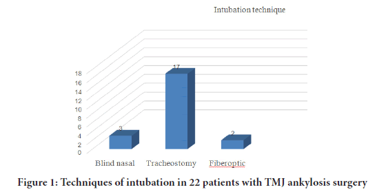
Tracheostomy was done in 17 (77%) as intubation technique, Blind nasal intubation was done in 3 (14%) and fiber optic was used only in 2 (9%) patients (Figure 1).

Figure 1: Techniques of intubation in 22 patients with TMJ ankylosis surgery
Temporomandibular Joint ankylosis is fusion of head of mandibular condyle to the glenoid fossa of temporal bone and it is a common disorder of the Temporomandibular Joint. This study was based on retrospective chart review in 22 patients who had been operated for TMJ ankylosis from January 2017 to December 2019. In this study, the distribution of males and females were equal 11 (50%). In contrast to this study, male predominance was reported in previous studies (El-Hakim IE and Metwalli SA, 2002; Punjabi SK, et al., 2013; Bello SA, et al., 2012).
The age range from 6-50 years with mean of 21.7 years and the frequency of TMJ ankylosis was highest in second decade (11-20 years) 9(40.91%) followed by third decade (21-30 years) which account for 7(31.82%). It may be due injury to TMJ in growing population remains unreported due to lack of facilities, trained professionals in diagnosis and lack of awareness subsequently leading to ankylosis. The finding of most frequently occurring age group was the same with the study done in Pakistan and Egypt as reported second decade was the most frequent age group (Hossain MA, et al., 2014; Elgazzar RF, et al., 2010).
In this study, the frequency of unilateral ankylosis was more predominant 13(59.09%) than bilateral ankylosis. This result was in line with the finding of other literatures (Vasconcelos BC, et al., 2011; Mabongo M and Karriem G, 2014). It was found that bony ankylosis reported in majority 15 (68.18%) of cases. However, this number was still less when compared to other studies (Tefera T, 2019). This difference could be accounted for by variations in types and mechanism of injury, age of patient and duration of ankylosis during diagnosis.
Intubation technique
Administration of anesthesia to patients with TMJ ankylosis poses specific problems and demands expertise. In current study, tracheostomy was done in majority of patients 17(77%) as means of intubation technique and only 2(9%) patients were intubated using fiberoptic. In the study conducted in South Africa fiberoptic was used in (50%) of patients and tracheostomy was done only in (17.5%) cases. The use of fiberoptic in this study is much less when compared to the study conducted in South Africa (Mabongo M and Karriem G, 2014). Blind awake intubation was successful in majority 92% of the cases done in India (Sankar D, et al., 2016), which is completely incomparable with our study finding only 14% were intubated by blind nasal. This lowest number of fibroptic and blind nasal intubation in our current study may be due an availability of fibroptic and lack of skilled anesthesiologist in practice of these techniques. Fiberoptic intubation is considered as a gold standard in TMJ anklosis surgery as means of airway securing. Tracheostomy is an invasive procedure with a post-operative morbidity (Shaeran TA and Samsudin AR, 2019).
Conclusion
Even though fibroptic is the golden standard for TMJ ankylosis surgery, in our study we found that tracheostomy was the most commonly used intubation technique, due to unavailability of fibroptic in the institutions and few patients 2(9%) operated on campaign intubated using fibroptic by team of maxillofacial surgeons from Canada at St. Peter specialized Hospital.
However, tracheostomy is an invasive procedure with a post-operative morbidity. Institutional capacity building of facilities; increasing service availability and trained professionals in practice of fibroptic and blind nasal intubation techniques is recommended. The anesthesia department should establish guidelines or algorithm specific to their institution depending on the expertise and the facility available in regard to TMJ anyklosis surgery.
Acknowledgement
My special thanks to my wife Chaltu Temesgen, would not have done this without her support and my little kids kaya and Marin.
References
- Al-Rawee RY, Al-Khayat AM, Salim Saeed S. True bony TMJ ankylosis in children: Case report. Int J Surg Case Rep. 2019; 61: 67-72.
- Miloro M, Ghali G, Waite P. Peterson's principles of oral and maxillofacial surgery. PMPH-USA. 2012.
- Kumar N, Sardana R, Kaur R, Jain A. Intraoperative mandibular nerve block with peripheral nerve stimulator for Temporomandibular Joint ankylosis. J Clin Anesth. 2016; 35: 207-209.
- Alyami YD, Abdulaziz H, Al T, Ramadan RM, Subahi JH, Haroon M, et al. Overview of Management of Temporomandibular Joint Ankylosis. Ec Dental Science. 2020;2:1-5.
- Yan YB, Liang SX, Shen J, Zhang JC, Zhang Y. Current concepts in the pathogenesis of traumatic Temporomandibular Joint ankylosis. Head Face Med. 2014; 10(1): 1-2.
- El-Hakim IE, Metwalli SA. Imaging of Temporomandibular Joint ankylosis: A new radiographic classification. Dentomaxillofacial Radiol. 2002; 31(1): 19-23.
- Kumar R, Hota A, Sikka K, Thakar A. Temporomandibular Joint ankylosis consequent to ear suppuration. Indian J Otolaryngol Head Neck Surg. 2013; 65(3): 627-630.
- Braimah RO, Taiwo AO, Ibikunle AA, Oladejo T, Adeyemi M, Adejobi AF, et al. Temporomandibular Joint Ankylosis with maxillary extension: Proposal for modification of Sawhney's classification. Craniomaxillofacial Trauma Reconstr Open. 2018; 2(1): 38.
- Güven O. A clinical study on Temporomandibular Joint ankylosis. Auris Nasus Larynx. 2000; 27(1): 27-33.
- Xia L, An J, He Y, Xiao E, Chen S, Yan Y, et al. Association between the clinical features of and types of Temporomandibular Joint ankylosis based on a modified classification system. Sci Rep. 2019; 9(1): 1-7.
- Potdar S, Devadoss V, Choudary N, Tiwari R, Pandey P, Mahajan S. Epidemiological study of Temporomandibular Joint ankylosis cases in a tertiary center. Int J Appl Dent Sci. 2019; 5: 142-145.
- Shirani G, Arshad M, Mahmoudi X. A new method of treatment of Temporomandibular Joint ankylosis with osteodistraction using the Sh-device: A case report. J Dent. 2018; 15(1): 63.
- Hossain MA, Shah SA, Biswas RS. Frequency of Temporomandibular Joint ankylosis in various age groups with reference to etiology. Chattagram Maa-O-Shishu Hosp Med Coll J. 2014; 13(2): 17-20.
- Gupta VK, Mehrotra D, Malhotra S, Kumar S, Agarwal GG, Pal US. An epidemiological study of Temporomandibular Joint ankylosis. Natl J Maxillofac Surg. 2012; 3(1): 25.
- Shaeran TA, Samsudin AR. Temporomandibular Joint ankylosis leading to obstructive sleep apnea. J Craniofac Surg. 2019; 30(8): 714-717.
- Wahal R. Temporo-Mandibular Joint ankylosis-The difficult airway. J Oral Biol Craniofacial Res. 2015; 5(2): 57.
- Jaju SR, Chhatrapati S, Ubale P, Ingle S. Anaesthetic management of a paediatric patient with bilateral Temporomandibular Joint ankylosis: A case report. Int J Contemp Med Res. 2018; 5(4).
- Punjabi SK, Channar KA, Munir A. A comparative study of gap and interpositional arthroplasty with temporalis myofacial flap for TMJ ankylosis treatment. Pak Oral Dental J. 2013; 33(3).
- Bello SA, Olokun BA, Olaitan AA, Ajike SO. Aetiology and presentation of ankylosis of the Temporomandibular Joint: Report of 23 cases from Abuja, Nigeria. Br J Oral Maxillofac Surg. 2012; 50(1): 80-84.
- Elgazzar RF, Abdelhady AI, Saad KA, Elshaal MA, Hussain MM, Abdelal SE, et al. Treatment modalities of TMJ ankylosis: Experience in Delta Nile, Egypt. Int J Oral Maxillofac Surg. 2010; 39(4): 333-342.
- Vasconcelos BC, Carvalho RW, Porto GG, Bessa-Nogueira RV, Nascimento MM. Surgical treatment of Temporomandibular Joint ankylosis: Follow-up of 15 cases and literature review. Int J Oral Maxillofac Surg. 2011; 40(10): 1224.
- Mabongo M, Karriem G. Temporomandibular Joint ankylosis: Evaluation of surgical outcomes. J Dent Med Sci. 2014; 13(8): 60-66.
- Tefera T. Incidence, clinical presentation and surgical management of Temporomandibular Joint ankylosis: A 5 year retrospective study. Scientific Archives of Dental Sciences. 2019; 2: 2-14.
- Sankar D, Krishnan R, Veerabahu M, Vikraman BP, Nathan JA. Retrospective evaluation of airway management with blind awake intubation in Temporomandibular Joint ankylosis patients: A review of 48 cases. Ann Maxillofac Surg. 2016; 6(1): 54.
Author Info
Gelana Garoma1*, Demerew Dejene1 and Ajay Prakash22Department of Dentistry, Wachemo University, Hosaena, Ethiopia
Received: 23-Jun-2021 Accepted: 07-Jul-2021 Published: 14-Jul-2021
Copyright: This is an open access article distributed under the terms of the Creative Commons Attribution License, which permits unrestricted use, distribution, and reproduction in any medium, provided the original work is properly cited.
ARTICLE TOOLS
- Dental Development between Assisted Reproductive Therapy (Art) and Natural Conceived Children: A Comparative Pilot Study Norzaiti Mohd Kenali, Naimah Hasanah Mohd Fathil, Norbasyirah Bohari, Ahmad Faisal Ismail, Roszaman Ramli SRP. 2020; 11(1): 01-06 » doi: 10.5530/srp.2020.1.01
- Psychometric properties of the World Health Organization Quality of life instrument, short form: Validity in the Vietnamese healthcare context Trung Quang Vo*, Bao Tran Thuy Tran, Ngan Thuy Nguyen, Tram ThiHuyen Nguyen, Thuy Phan Chung Tran SRP. 2020; 11(1): 14-22 » doi: 10.5530/srp.2019.1.3
- A Review of Pharmacoeconomics: the key to “Healthcare for All” Hasamnis AA, Patil SS, Shaik Imam, Narendiran K SRP. 2019; 10(1): s40-s42 » doi: 10.5530/srp.2019.1s.21
- Deuterium Depleted Water as an Adjuvant in Treatment of Cancer Anton Syroeshkin, Olga Levitskaya, Elena Uspenskaya, Tatiana Pleteneva, Daria Romaykina, Daria Ermakova SRP. 2019; 10(1): 112-117 » doi: 10.5530/srp.2019.1.19
- Dental Development between Assisted Reproductive Therapy (Art) and Natural Conceived Children: A Comparative Pilot Study Norzaiti Mohd Kenali, Naimah Hasanah Mohd Fathil, Norbasyirah Bohari, Ahmad Faisal Ismail, Roszaman Ramli SRP. 2020; 11(1): 01-06 » doi: 10.5530/srp.2020.1.01
- Manilkara zapota (L.) Royen Fruit Peel: A Phytochemical and Pharmacological Review Karle Pravin P, Dhawale Shashikant C SRP. 2019; 10(1): 11-14 » doi: 0.5530/srp.2019.1.2
- Pharmacognostic and Phytopharmacological Overview on Bombax ceiba Pankaj Haribhau Chaudhary, Mukund Ganeshrao Tawar SRP. 2019; 10(1): 20-25 » doi: 10.5530/srp.2019.1.4
- A Review of Pharmacoeconomics: the key to “Healthcare for All” Hasamnis AA, Patil SS, Shaik Imam, Narendiran K SRP. 2019; 10(1): s40-s42 » doi: 10.5530/srp.2019.1s.21
- A Prospective Review on Phyto-Pharmacological Aspects of Andrographis paniculata Govindraj Akilandeswari, Arumugam Vijaya Anand, Palanisamy Sampathkumar, Puthamohan Vinayaga Moorthi, Basavaraju Preethi SRP. 2019; 10(1): 15-19 » doi: 10.5530/srp.2019.1.3


