Review Article - (2022) Volume 13, Issue 6
Abstract
Diuretic agents already prove its clinical affectivity against various disease conditions like hypertension, renal failure, nephritic syndrome etc. In this case, various in vitro and in vivo screening models express their clinical importance. The present review emphasis about rational purpose of experimental techniques and evaluation parameters of various screening models of diuretic activity. It also reveals the usefulness and importance of these screening techniques regarding the evaluation of safety and affectivity of different members from various classes of diuretic agents. This review also highlighted on effective several modifications of some screening models.
Keywords
Diuretic agent, in vivo techniques, in vitro screening models, Evaluation parameters, Modifications
Introduction
The term diuretic is originally derived from an ancient term, “Diu oyr1ih” (Diu means through and oyr1ih means to urinate). Diuretic concerns any material which promotes rate of urine formation and also facilities excretion of water. It primarily increases the excretion of various electrolytes like sodium (Na+), Chlorine (Cl-), Bicarbonate (HCO3) and water. It also inhibits reabsorption sodium (Na+), Chlorine (Cl-), Bicarbonate (HCO3) and water. So final outcome will be the i) increased rate of urine formation ii) altered Urine pH iii) ionic composition of Urine and blood will also be altered (Vogel GH, 2016).
Role of kidney in excretion
Kidney plays important role in excretion. Basic functional unit of kidney is Nephron. Pathophysiology of renal excretion system includes 3 main components like i) Glomerular filtration ii) Proximal Convoluted Tubule (PCT), Distal Convoluted Tubule (DCT) mediated reabsorption iii) active secretion. It is shown in Figure 1.
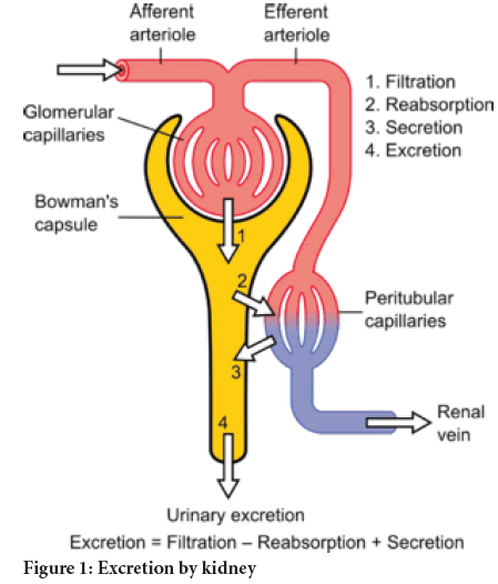
Figure 1: Excretion by kidney
Glomerular filtration receives 25% of cardiac output. The filtration rate is 80-100 ml/min. Normal per day capacity of filtration for Bowman’s capsule is 180 Lit. Proximal and Distal Convoluted Tubules mostly reabsorb glomerular filtrate. Proximal Convoluted Tubule reabsorbs 60%-70% of sodium. It is water permeable. It is isotonic with urine. It favors reabsorption of glucose, amino acid, cations. The prime target for water reabsorption is Proximal Convoluted Tubule. Loop of henle shows sodium reabsorption. It is water impermeable. Distal tubule favors sodium reabsorption. Collecting duct is water impermeable.
Rational mode of diuretics
Drugs belonging to diuretic class show desired effect through the inhibition of reabsorption of several electrolytes like potassium, sodium, Chlorine and bicarbonate etc. Loop of henle, early distal tubule, late distal tubule and collecting duct which are offer themselves as a prime target site for diuretics (Okusa MD and Ellison DH, 2008). The following Table 1 clearly exploits several classes of diuretic along with their site of action.
| Diuretics | Target sites | Effect on excretion |
|---|---|---|
| Osmotic Diuretic | Proximal convoluted tubules, Loop of Henle, Collecting duct | Increases excretion of sodium and water |
| Carbonic Anhydrase Inhibitor (CA-I) | Proximal tubules | Increases excretion of sodium bicarbonate |
| High ceiling loop.diuretic | Thick ascending loop of henle | Increases excretion of NaCl and KCl |
| Thiazides | Early distal convoluted tubule | Increases excretion of NaCl |
| K+ .sparing. diuretics | Late distal. tubule, Collecting duct | Increases excretion of Na+ and K+ secretion |
Table 1: Site and mechanism of action of different diuretics
Diuretic activity evaluating models
There are several in vivo and in vitro methods are available for evaluation of diuretic activity. Urine volume, electrolyte concentration these are common evaluating parameters in it (Takehito T, et al., 1991).
Literature Review
These are several methods for diuretic activity evaluation as follows:
1. in vitro methods
• Carbonic Anhydrase (CA) inhibition • Patch clamp technique in kidney cells
• Perfusion of isolated kidney tubules
2. in vivo methods
• Lipchitz value
• Saluretic activity in rats
• Diuretic and Saluretic activity in dogs
• Micro puncture techniques in rats
• Stop flow technique
In vitro methods
Carbonic Anhydrase inhibition:
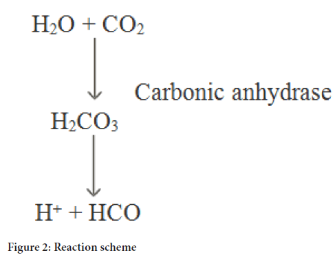
Rational background: The first recognized member from this class is Acetazolamide (Diamox®). It antagonizes activity of Carbonic Anhydrase enzyme. As a structural point of view, it is synthetic derivative and exhibits presence of zinc in their structure. It is mainly responsible for formation of carbonic acid from CO2 and water. It also carries reabsorption of sodium, bicarbonate and water in Proximal Convoluted Tubule. So these inhibiters blocks reabsorption of sodium, bicarbonate and water by antagonizing activity of enzyme Carbonic Anhydrase. In 1960, Scientist Maren TH was described micro method which sounds easy and efficient (Maren TH, 1960). Red cells are full enrichment for it. Another prime source of the same is enzymes also located in the eye (Frost SC and McKenna R, 2013; Carta F and Supuran CT, 2013). Reaction scheme is proceed as shown in Figure 2.
Figure 2: Reaction scheme
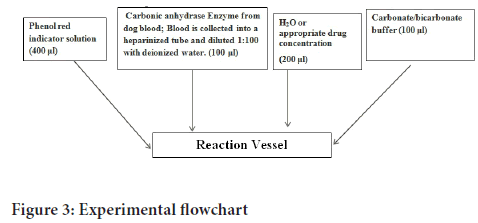
Experimental procedure: Reaction Vessel (Monostat bench mounted flowmeter) is used. CO2 flow rate is maintained on 30-45 ml/min. Experimental flow chart is shown in Figure 3 (Maren TH, 1960). In this model, following parameters are evaluated in duplicate samples:
Tu=(Uncatalyzed time)=required time to occur color change in the absence of enzyme.
Te=(Catalyzed time)=required time to occur color change in the presence of the enzyme.
Tu-Te=enzyme rate.
Ti=enzyme rate with the presence of assorted concentrations of inhibitor
Calculation: %reduction in activity of Carbonic Anhydrase enzyme is measured by employing below formula:
%Inhibition=1-[(Tu-Te)-(Ti-Te)/(Tu-Te)] × 100
Measurement of Percent inhibition of CA-inhibitors is effective tool to access the diuretic potential of several sulfonamides. There are several implementation have been reported for this procedure (Landolfi C, et al., 1997). It measures time which is required to for pH alternation between 8-7.5 and further alternation in time period and pH can be achieved with use of such CA enzyme inhibitors
Figure 3: Experimental flowchart
Patch clamp technique in kidney cells:
Principle ration: Entire excretion process includes the various segments of the kidney such as like loop of henle, early and late convoluted tubules etc. plays important role in fluid reabsorption. So in that case flow of substance either from the tubular lumen to the blood stream (i.e. tubular reabsorption) or active secretion. Apart from active transport, there are several coupled transport systems also available. Out of them, ion channels show strong influence over the function of kidney cells. Various types of this technique quite differ from each other with respect of use of single and whole cell ion channel. It allows a use of patch electrode consisting relatively large tip (greater than 1 mm) along with smooth surface.
There are various technique modes of patch clamp as follow,
1. Attached mode with cell
2. Excised mode with cell
3. whole-cell mode
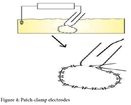
Experimental procedure: Experimental technique allows compression of patch-clamp electrode opposite to a cell membrane and vacuum is exerted to insert the cell membrane within the tip of electrode.
Due to such vacuum pressure cell creates tight, high-resistance gigaseal with electrode, ( ≥ 10 giga Ohms), as shown in Figure 4 (Burg M, et al., 1966).
Figure 4: Patch-clamp electrodes
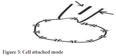
Cell-attached mode: This technique exhibits sealing the patch electrode within the cell membrane, which allows the passage of currents through from the membrane patch within vicinity single-ion channels which is covered by electrode tip (as shown in Figure 5).
Figure 5: Cell attached mode
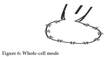
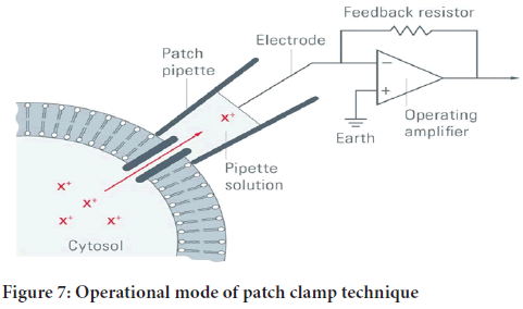
Whole-cell mode: While comparison to previous cell-attached mode, extra more suction is employed through which rupture of the cell membrane is achieved, so the inner cell passage experiences more access to it. The electrode’s content takes a place of cell’s soluble content. This technique permits passage of currents from entire ion channels of whole membrane in a single operation only (as shown in Figure 6 and operational technique is represented in Figure 7).
Figure 6: Whole-cell mode
Figure 7: Operational mode of patch clamp technique
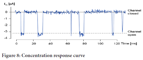
Evaluation: A graph of drug’s concentration vs. ion channel inhibition is plotted. Whole cell operational mode from patch clamp technique provides better and effective estimation of sodium- Alanine co transport with help of separated cell from ascending loop of henle. The apparent Km values for sodium and L-alanine can be recorded (as shown in Figure 8).
Figure 8: Concentration response curve
Modifications: Several modifications have been made by Scientist using voltage gated macula densa cells along with whole-cell operational mode from patch-clamp technique. In this technique effect of diuretics as well as voltage gated ion potential of macula densa cell were effectively estimated.
Perfusion of isolated kidney tubules:
Rational principle: The different tubules fractions like thin ascending loop of henle, Distal Convoluted Tubules etc. have different functional properties. If the target site and Mode of action of diuretics is clearly known (mostly regarding the clearance and micro puncture studies) at that time this is method of choice. This technique involves measurement of change in concentration of solutes in perfusion fluid (Schafer JA and Andreoli TE, 1979).
Procedure: In 1966, Scientist Burg and his coworker were invented above perfusion mode of isolated kidney tubules. After his invention, it has been effectively utilized with different animal species e.g. Wrister albino rat, mice, rabbit, etc. The thin (<1 mm) tubule fragments are isolated from kidney and afterwards subjected into development assembly. To perfuse suitable tubule, one end of the tubule is holed by micropipette. A perfusion pipette is immersed into lumen of kidney. The remaining end of the tubule is sucked into collecting pipette. The oil inside the collecting pipette prevents the evaporation. Entire gathered fluid is received at suitable time intervals by immersing a narrow gradual pipette in the collecting pipette. To approximate in vivo situation, an isotonic rabbit serum sample is collected by inserting tubule in rabbit serum bath (as shown in Figure 9) (Burg MB, 1982).
Figure 9: Tubule with perfusion pipette
Evaluation: The absolute volume of reabsorption is determined from the change in the concentration of an impermeable marker like (3H) insulin, (125I) isothalamate in the collecting fluid. The presence of leaks around the perfusion pipette is detected from the appearance of the marker in the external bath (Jacobson HR and Kokko JP, 1982).
In vivo methods
Lipchitz test:
Principle: Lipchitz value measures ratio of water and sodium excretion of test.animals (rats) to that of rats which are already treated with Standard drug (dose as per reference) (Lipschitz WL, et al., 1943).
Methodology: Species: Wistar albino rats (100-200 gm)
Sex: Male/female rats
Animals are divided into 3 groups like as a test, control and standard (6 animals in each group).
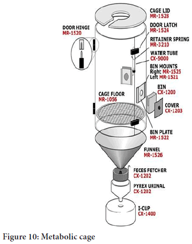
Procedure: In this procedure animals are divided into 5 groups. Each group contains 6 animals. Animals from each group are placed in metabolic cages which are designed in such way that wire mesh bottom and funnel for easy collection of the urine. Funnel is maintained with SS sieves to restrict feces but only passes urine. Group I receives control (e.g. normal saline solution), Group II receives reference standard (in standard dose as per reference) while Group III, IV, and V are received test sample in dose according to acute toxicity study (mild, moderate, high). Before experimentation, visible signs of disease are screened in animals and only healthy animals are allowed for experiment. Food and water is withdrawn before 17-24 hrs of experiment. During study a normal room temperature (25 ± 2OC) is maintained. Prime care has to be taken that before dosing of sample/controls; Rat’s bladder is become empty by exerting pressure on pelvic area and by stretching of tails. It ensures same quantity of dose is administered in each animal, which are mostly made in equal volume of normal saline. This procedure mostly intraperitoneal route is preferred for administration of reference/test which provides easiness and safety in concern of administration of larger doses of fluid. After administration, animals are subjected to metabolic cage which is specially designed to collect urine. After 5 and 24 hrs, Urine excretion volume is recorded. Flame photometry is used to determine Na content of urine. Lower doses are preferred to test active compound (as shown in Figure 10) (Sathianarayanan S, et al., 2011).
Figure 10: Metabolic cage
Evaluation: Following formula is employed to measure this index.
Lipchitz value=Urine output in test/Urine output in Standard
Results are mentioned according to following predictions of this value which are represented in above Table 2.
| Lipschitz value | Diuretic activity |
|---|---|
| Greater than 1 | Positive impact |
| Greater than 2 | Potent impact |
Table 2: Interpretation of activity
Saluretic activity in rats:
Rational principle: The peripheral edema, congestive heart failure and hypertension are treated by maintain excretion of electrolyte and water. In that case however potassium (K+) loss has to be avoided so from this incidence, there is development of Saluretic and Potassium sparring diuretic occurred. There are several modifications has been made in this diuretic test of rat in such way that along with sodium and water content determination it also includes determination of potassium and Chlorine (Cl-) content. Osmolality determination is also part of this test. Carbonic Anhydrase (CA) inhibition and potassium sparring effect is estimated through calculation of electrolyte ratio (Bicking JB, et al., 1965).
Experimental technique: This test can be performed with mostly Wister rat species (100-200 g) of male sex only. Animals are divided into 3 groups as like Control, Standard and Test. Each group contains 3 animals. Male Wistar rats are maintained on normal diet like Altromin pellets and water. Before 15.hrs of experiment only food is withdrawn. Test compounds are administered through oral route of dose 50 mg/kg in 0.5 ml/100 g body weight of starch suspension. For proper purpose of urine collection, 3 animals are placed each metabolic cage should carries only 3 animals along with wire mesh and funnel assembly at bottom. Urine excretion volume is measured at 1 hr interval up to 5 hrs. After 5 hrs, Sodium (Na+) and Potassium (K+) content from collected urine sample is estimated by using Flame Photometry. Prolonged effect of test sample is estimated by analyzing urine sample which is collected up to 24 hrs. In this technique Furosemide or Hydrochlorothiazide is preferred as a standard (Kagawa CM, et al., 1957).
Evaluation: Saluretic activity is measured through sum of sodium and chloride excretion (Na+ excretion+Cl- excretion). Natriuretic activity is calculated through ratio of Sodium excretion to that of Potassium excretion (Na+ excretion/K+ excretion). Carbonic Anhydrase inhibition is calculated through ratio of chloride excretion to that of sum sodium and potassium excretion (Cl- excretion/Na+ excretion+K+ excretion). This ratio is known as ion quotient. Activity interpretation is as shown in Table 3.
| Activity | Value |
|---|---|
| Natriuretic effect | Values>2 |
| K sparing effect | Ratios>10 |
Table 3: Interpretation of activity
Modification: Several modifications have reported in this method in concern of Aldosterone antagonists study. It can be conducted by using Adrenolectomized Rats thoughts are treated with aldosterone.
Diuretic and Saluretic activity in dogs:
Rational principle: In comparison to rats, dog’s renal physiology is supposed to be quite similar to man. So dogs are considered more appropriate to study oral absorbability of diuretic substances. Urine collections at respective time period interval can become a simple and compatible with use of catheters. Pharmacokinetics properties are also studied by withdrawing blood samples.
Experimental Technique: For this study dogs (male/female) are used. Animals are divided into three groups (Control, Reference, and Test). Each group contains four animals. Intensive training to be accustomed for Beagle dogs of either sex have to gain gavages feeding and catheterization after every hr. During this period primly monitored for any sign of resistance. Mostly metabolic cages were preferred for it. Water is used as Control While, as standard controls either urea (1 g/kg, po) or furosemide (5 mg/kg, po). Before 24 hrs of experiment only food is withdrawn (water supply maintains). Urinary bladder is made empty by using plastic catheter especially on the morning of the experiment. Dogs were received 20 ml/kg water through gavages, followed by potable dose of 4 ml/kg at 1 hr interval. Initial values are analyzed by catheterizing bladder for two times at 1 hr interval and afterwards urine sample was collected. Either oral or intravenous route is preferred for both test and standard sample. Hourly catheterization is perennial over consequent six hrs. Animals are subjected to metabolic cages for longer period of time so additional water supply was maintained. The dogs are catheterized once more only after 24 hrs of dosage of the test compound and keep record of urine volume along with together with the urine collected over night collected urine in the metabolic cage. Electrolyte concentration like Na+ and K+ are analyzed by using flame photometer and Cl- contents are analyzed with use of Argentometry. Furthermore, osmolality evaluation can be done with an Osmometer.
Observations: The common evaluating parameters are like urine output, electrolyte content and glomerular filtration rate. Comparison of these pretreatment values with water, controls and standards can be done by plotting against time. Statistical analysis can be performed by using non-parametric U-test.
Micro puncture techniques in rats:
Rational principle: Micro puncture techniques discovered influence of diuretic on nephron activity. The changes in rate of tubular fluid reabsorption and electrolyte concentrations are mainly used as evaluating parameters to confirm effectivity of this technique. This technique is mostly conducted with rat. The Prime target sites for micro puncture action are thick ascending loop of henle, Distal Convoluted Tubules and collecting ducts also (Shipp JC, et al., 1958).
Experimental technique: This technique is performed with rats (250 g body wt) of either sex. Thiopentone injection is administered through intraperitoneal route in rats for anesthesia purpose. Before start of the experiment, only food is withdrawn but allows water access. Only after anesthesia, the animals are maintained on a thermostatically heated table and then after rats are tracheotomized. BP (Blood Pressure) measurement, blood sample collection, and compounds remedy such functions are conducted through cannulation of carotid artery and jugular vein. Flank incision allows access of retroperitoneum region (excretory organ), which get enclosed in a small plastic object along with cotton, and finally soaked with liquid paraffin oil at room temp. Cannula is inserted into ureter and body temperature observed incessantly. After 3 hrs, single large dose of inulin injection which is prepared in NaCl solution and directly administered into bolus, then immediately 0.85% NaCl solution is administered with flow rate of 2.5 ml/ min and dose is calculated suitably as per 100 g body weight. intravenous infusion is creates a gradual leakage of slender,elongated channel only after 45 min. Glass capillaries (8 to10 μm external diameter) is used to directly collect intracellular liquid sample from renal cortex and medulla. A micromanipulator and microscopic observation is used for it. Lissamine green (through Intravenous route) is used to identify distal tubule. Test samples are administered only after control sample. Micro puncture is performed again only after 1/2 hr equilibration period which get attained along with sample specimen then collect tubular fluid. Urethral urine and blood sample is collected between clearance periods (Ulrich KJ, et al., 1969).
Evaluation: The evaluating parameters are as follow-
1. Inulin clearance (GFR)
2. Single nephron GFR
3. Fractional delivery of water
4. Sodium and potassium electrolyte concentration in renal tubules and in urine also.
The statistical techniques like one-way Analysis of Variance (ANOVA) and Student’s t-test are applied to measured values of evaluating parameters to compare between paired and unpaired data. Results are exploited as mean values ± SEM (Standard Error of the Mean).
Stop flow technique:
Rationale principal: The passages of renal tubular fluid which get lined along with nephron structure are considerably localized by using stop flow technique. Glomerular filtration rate is significantly reduced during clamping of the ureter. When electrolyte concentration of intracellular renal sample specimen is in ultimate static equilibrium-head condition, at that time it increase contact time between tubular fluid and respective nephron segments. Once clamp is released, it slightly modifies composition of the tubular fluid. The first sample is collected from correspond the distal nephron segment and last followed from glomerular fluid. Now days, stop-flow method is considered to be least preferred method in comparison with micro puncture technique (Tobian L, et al., 1964).
Experimental methodology: Stop flow technique can be conducted with different animal species. This technique allows clamping of animal’s ureter for several minute which generates intense osmotic dieresis. Due to it, equilibrium renal column pressure permits intact exposure between different renal segments for extra time period in comparison with normal time period. So, experimental technique on every segment of tubular fluid is enlarged. Once clamp is released then urine sample is collected, respectively. In this series of activity, smaller samples are collected in rapid manner while earlier collected samples expressing tubular fluid which is in contact with distal part of nephron. Test samples are administered along with inulin before occlusion of urethra is carried out. Downstream content of tubules especially from proximal part of nephron may alters the tubular fluid content at time of enlargement (Nagaoka Y, et al., 2018).
Evaluation:Stop flow technique measures concentration content glomerular marker sample such as inulin along concentration content of test samples. After that fractional excretion volume of marker and test sample is plotted against cumulative urinary volume.
Modifications: Several stop flow studies reported which are conducted with tubular secretion inhibitor as like pyrazinoic acid especially on uric acid transport in rats. Scientist Nagaoka Y, et al. is used this technique on dogs to study the effect of uricosuric drugs also (Nagaoka Y, et al., 2018).
Conclusion
Now days several efficient screening techniques are available to evaluate diuretic efficiency of diuretic agents. It is most effective way to judge potency as well as efficacy of various diuretic agents. It also provides useful information regarding level of dosage regimen criteria of particular class of diuretic agents. Modification reveals recent advances in existing technique. It also reveals the usefulness and importance of these screening techniques regarding the evaluation of safety and effectivity of different members from various classes of diuretic agents.
References
- Vogel GH. Drug discovery and analysis medicine Assay. Springer. 2016; 317-348.
- Okusa MD, Ellison DH. Physiology and pathophysiology of diuretic action. Seldin and Giebisch's the kidney. 2008; 1051-1094. [Crossref]
- Takehito T, Kazuyo N, Tsuyoshi K, Yoshifumi M, Masataka M, Eiji S. Validation of a toxicity testing model by evaluating oxygen supply and energy state in the isolated perfused rat kidney: Single-pass preparation without albumin. J Pharmacol Methods. 1991; 25(3): 195-204. [Crossref]
[Google Scholar] [Pubmed]
- Maren TH. A simplified micromethod for the determination of carbonic anhydrase and its inhibitors. J Pharmacol Exp Ther. 1960; 130(1): 26-29.
[Google Scholar] [Pubmed]
- Frost SC, McKenna R. Carbonic anhydrase: Mechanism, regulation, links to disease, and industrial applications. Springer Science and Business Media. 2013.
- Carta F, Supuran CT. Diuretics with carbonic anhydrase inhibitory action: A patent and literature review (2005-2013). Expert Opin Ther Pat. 2013; 23(6): 681-691. [Crossref]
[Google Scholar] [Pubmed]
- Landolfi C, Marchetti M, Ciocci G, Milanese C. Development and pharmacological characterization of a modified procedure for the measurement of carbonic anhydrase activity. J pharmacol Toxicol Methods. 1997; 38(3): 169-172. [Crossref]
[Google Scholar] [Pubmed]
- Burg M, Grantham J, Abramow M, Orloff J. Preparation and study of fragments of single rabbit nephrons. Am J Physiol. 1966; 210(6): 1293-1298. [Crossref]
[Google Scholar] [Pubmed]
- Schafer JA, Andreoli TE. Perfusion of isolated mammalian renal tubules. Transport Organs. 1979; 473-528. [Crossref]
- Burg MB. Introduction: Background and development of microperfusion technique. Kidney Int. 1982; 22(5): 417-424. [Crossref]
[Google Scholar] [Pubmed]
- Jacobson HR, Kokko JP. Initial remarks and acknowledgments: Isolated perfused tubule symposium. Kidney Int. 1982; 22(5): 415-416. [Crossref]
[Google Scholar] [Pubmed]
- Lipschitz WL, Hadidian Z, Kerpcsar A. Bioassay of diuretics. J Pharmacol Exp Ther. 1943; 79(2): 97-110. [Crossref]
- Sathianarayanan S, Jose A, Rajasekaran A, George RM, Chittethu AB. Diuretic activity of aqueous and alcoholic extracts of Wrightia tinctoria. Phytomedicine. 2011; 2: 7-8.
- Bicking JB, Mason JW, Woltersdorf Jr OW, Jones JH, Kwong SF, Robb CM, et al. Pyrazine diuretics. I. N-amidino-3-amino-6-halopyrazinecarboxamides. J Med Chem. 1965; 8(5): 638-642. [Crossref]
[Google Scholar] [Pubmed]
- Kagawa CM, Cella JA, van Arman CG. Action of new steroids in blocking effects of aldosterone and deoxycorticosterone on salt. Science. 1957; 126(3281): 1015-1016. [Crossref]
[Google Scholar] [Pubmed]
- Shipp JC, Hanenson IB, Windhager EE, Schatzmann HJ, Whittembury G, Yoshimura H, et al. Single proximal tubules of the Necturus kidney: Methods for micropuncture and microperfusion. Am J Physiol. 1958; 195(3): 563-569. [Crossref]
[Google Scholar] [Pubmed]
- Ullrich KJ, Frömter E, Baumann K. Micropuncture and microanalysis in kidney physiology: Laboratory techniques in membrane biophysics. 1969; 106-129. [Crossref]
- Tobian L, Coffee K, Ferreira D, Meuli J. The effect of renal perfusion pressure on the net transport of sodium out of distal tubular urine as studied with the stop-flow technique. J Clin Invest. 1964; 43(1): 118-128. [Crossref]
[Google Scholar] [Pubmed]
- Nagaoka Y, Tanaka Y, Yoshimoto H, Suzuki R, Ryu K, Ueda M, et al. The effect of small dose of topiroxostat on serum uric acid in patients receiving hemodialysis. Hemodial Int. 2018; 22(3): 388-393.
[Crossref] [Google Scholar] [Pubmed]
Author Info
Kiran M Kulkarni* and Manish S KondawarCitation: Kulkarni KM: Review on Emerging Trends and Modifications of In vivo and In vitro Screening Techniques of Diuretic Activity
Received: 02-May-2022 Accepted: 27-May-2022 Published: 03-Jun-2022, DOI: 10.31858/0975-8453.13.6.399-404
Copyright: This is an open access article distributed under the terms of the Creative Commons Attribution License, which permits unrestricted use, distribution, and reproduction in any medium, provided the original work is properly cited.
ARTICLE TOOLS
- Dental Development between Assisted Reproductive Therapy (Art) and Natural Conceived Children: A Comparative Pilot Study Norzaiti Mohd Kenali, Naimah Hasanah Mohd Fathil, Norbasyirah Bohari, Ahmad Faisal Ismail, Roszaman Ramli SRP. 2020; 11(1): 01-06 » doi: 10.5530/srp.2020.1.01
- Psychometric properties of the World Health Organization Quality of life instrument, short form: Validity in the Vietnamese healthcare context Trung Quang Vo*, Bao Tran Thuy Tran, Ngan Thuy Nguyen, Tram ThiHuyen Nguyen, Thuy Phan Chung Tran SRP. 2020; 11(1): 14-22 » doi: 10.5530/srp.2019.1.3
- A Review of Pharmacoeconomics: the key to “Healthcare for All” Hasamnis AA, Patil SS, Shaik Imam, Narendiran K SRP. 2019; 10(1): s40-s42 » doi: 10.5530/srp.2019.1s.21
- Deuterium Depleted Water as an Adjuvant in Treatment of Cancer Anton Syroeshkin, Olga Levitskaya, Elena Uspenskaya, Tatiana Pleteneva, Daria Romaykina, Daria Ermakova SRP. 2019; 10(1): 112-117 » doi: 10.5530/srp.2019.1.19
- Dental Development between Assisted Reproductive Therapy (Art) and Natural Conceived Children: A Comparative Pilot Study Norzaiti Mohd Kenali, Naimah Hasanah Mohd Fathil, Norbasyirah Bohari, Ahmad Faisal Ismail, Roszaman Ramli SRP. 2020; 11(1): 01-06 » doi: 10.5530/srp.2020.1.01
- Manilkara zapota (L.) Royen Fruit Peel: A Phytochemical and Pharmacological Review Karle Pravin P, Dhawale Shashikant C SRP. 2019; 10(1): 11-14 » doi: 0.5530/srp.2019.1.2
- Pharmacognostic and Phytopharmacological Overview on Bombax ceiba Pankaj Haribhau Chaudhary, Mukund Ganeshrao Tawar SRP. 2019; 10(1): 20-25 » doi: 10.5530/srp.2019.1.4
- A Review of Pharmacoeconomics: the key to “Healthcare for All” Hasamnis AA, Patil SS, Shaik Imam, Narendiran K SRP. 2019; 10(1): s40-s42 » doi: 10.5530/srp.2019.1s.21
- A Prospective Review on Phyto-Pharmacological Aspects of Andrographis paniculata Govindraj Akilandeswari, Arumugam Vijaya Anand, Palanisamy Sampathkumar, Puthamohan Vinayaga Moorthi, Basavaraju Preethi SRP. 2019; 10(1): 15-19 » doi: 10.5530/srp.2019.1.3


